Intraocular Laser Photocoagulation Saves the Eye of a Patient with Severe Chemical Burns
While at work, a patient accidentally had hot chemicals splashed into his eye, placing him at high risk of severe vision loss. Doctors from the Department of Ophthalmology at Military Hospital 103 (Vietnam Military Medical University) successfully applied intraocular laser photocoagulation, helping to stabilize intraocular pressure and preserve optimal conditions for subsequent reconstructive treatment.
On December 13, the Department of Ophthalmology at Military Hospital 103 announced that its medical team had successfully performed endoscopic cyclophotocoagulation (ECP) with intraocular camera assistance to control secondary glaucoma in a patient suffering from total corneal fibrosis resulting from severe ocular burns.
The patient, Bui Hung Son, from Thai Nguyen Province, was transferred to the hospital on June 3, 2025, in critical condition, presenting with extensive corneal damage and a high risk of requiring enucleation. Thanks to timely emergency management, the eyeball was preserved and the intraocular structures were maintained, providing a foundation for subsequent treatment.

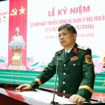
Colonel Dr. Nguyen Le Trung, Deputy Head of the Department of Ophthalmology, examines the patient’s eye following treatment with intraocular laser photocoagulation.
During follow-up, the patient developed secondary glaucoma, causing persistent pain and threatening the remaining function of the eye. Because the cornea had become completely opaque and the conjunctival fornices were severely shortened, conventional methods of intraocular visualization were not feasible. In this situation, the ophthalmology team decided to employ ECP, using an intraocular camera to directly access the ciliary body, clearly visualize the ciliary processes, and perform precise laser photocoagulation.
To facilitate the procedure, the patient underwent vitrectomy and removal of the opacified crystalline lens, thereby expanding the intraocular space to allow insertion of the camera and laser probe. Intraoperative findings revealed significant technical challenges due to total corneal fibrosis and severe symblepharon, which limited surgical instrument maneuverability. Nevertheless, the procedure was performed safely and successfully. Postoperatively, intraocular pressure was effectively controlled, and the posterior segment structures were preserved, creating favorable conditions for future visual rehabilitation, including corneal transplantation when circumstances permit.
Sharing his experience, the patient expressed his satisfaction, noting that throughout his treatment at the Department of Ophthalmology, doctors and medical staff provided attentive and compassionate care, treating him like a family member. This support helped him remain optimistic and contributed to his rapid stabilization.
According to Colonel Dr. Nguyen Le Trung, Deputy Head of the Department of Ophthalmology, the application of ECP at Military Hospital 103 significantly expands treatment options for patients with severe corneal damage who cannot be managed using conventional approaches. The success of this technique not only stabilizes intraocular pressure but also preserves optimal conditions for subsequent reconstructive interventions, including corneal transplantation, with the ultimate goal of restoring vision in the future.
In the coming period, the Department of Ophthalmology will continue to update, develop, and apply advanced techniques to meet increasingly demanding treatment requirements and to effectively fulfill its mission of protecting the health of military personnel and the general public.
News and images: Tuan Dung, Translator: Le Vien Lan Huong